Would a venous blood gas give the required information?
INDICATIONS
- Moderate or severe respiratory failure
- Patients with severe respiratory or cardiac disease scheduled for major abdominal or thoracic surgery
- Suspected acid-base disturbance
- Suspected carbon monoxide poisoning
- Emergency blood sampling when venepuncture impossible
WHICH ARTERY?
Radial Artery
Position of patient
- Arm extended and supported on pillow with wrist extended 20 º
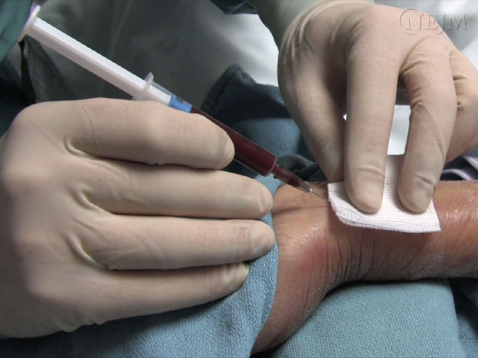
Angle of needle to skin
- 30º. See figure 1
Puncture site
- Proximal to proximal transverse crease on radial aspect of wrist
Advantages
- Easily accessible
- Easily compressible, therefore useful if there is known bleeding tendency
Contraindications
- Buerger's disease
- Raynaud's disease
- Arteriovenous dialysis shunt present or imminent
- Absent ulnar collateral circulation – relative contraindication, consider Allen’s test
Brachial Artery
Position of patient
- Arm extended and supported on pillow
Angle of needle to skin
- 30º
Puncture site
- Medial to biceps tendon in antecubital fossa
- CARE: Median nerve medial
Advantages
- Easily accessible
Disadvantages
- Risk of ischaemia
Contraindications
- Arteriovenous fistula in arm
- Elbow fractures
Femoral Artery
Position of patient
- Supine
Angle of needle to skin
- 60º
Puncture site
- Mid-inguinal point 2 cm below inguinal ligament
- CARE: Femoral nerve lateral and femoral vein medial
Advantages
- May be only quickly accessible artery in shocked patient
Disadvantages
- Risk of infection and ischaemia
- Venous sample more likely than at other sites
Contraindications
- Severe peripheral vascular disease
- Aortofemoral bypass surgery
EQUIPMENT
- Non-sterile disposable gloves
- Alcohol wipes or other antiseptic solution
- Lidocaine 1% plain 2 mL, 25 or 27 G needle and/or ice pack
- Blood gas syringe with 23 G needle
- smaller needles have shown longer draw times, and no pain benefit
- Plastic syringe cap
- Cotton wool balls or similar to press over site after arterial puncture
- Sharps' Bin
PROCEDURE
- If not competent in procedure, organise supervision by a clinician experienced in the procedure
Consent
- Explain procedure and reassure patient
- Obtain and record consent
- Positive Patient Identification (PPID) confirmed
Preparation
- If blood gas analysis not going to be performed within a few minutes, have an ice bag ready to cool sample
- Consider using ice (in a plastic bag) on skin for up to 3 min or cryogesic spray for additional/alternative analgesia to lidocaine
- Check concentration of oxygen patient is breathing at time arterial sample is taken
- if time permits, check it remains constant for 15 min before sampling
- note it on request form, in patient notes and on results printout
- Note patient’s temperature on request form
Aseptic technique and position of patient
- Select site of puncture and position patient. See WHICH ARTERY
- Wear gloves, cleanse patient’s skin
Local anaesthetic
- Palpate artery and infiltrate skin with lidocaine plain 1% 0.5–1 ml
- always aspirate before injection of local anaesthetic to prevent injection of lidocaine into the artery
Sampling
- Hold blood gas syringe with 23 G needle, bevel up; for radial (Figure 1) and brachial arteries at about 30° to skin surface; for femoral artery at 60°
- Advance needle towards artery
- with some blood gas syringes, blood pulsates into syringe; others will need to be drawn
- if shooting pain felt, nerve may have been entered. Remove needle and redirect
- if no blood obtained, withdraw needle slowly, observing for pulsation at base of needle; arterial blood often enters during withdrawal
- if necessary, try once more. If unsuccessful, seek help
- Obtain 1.5–2 mL blood
- a smaller volume may suffice for immediate analysis
- Withdraw needle
- Apply pressure to site for 5 min, or if site bleeds, longer
- Dispose of needle in sharps bin
- Remove bubbles in syringe by holding hub upwards and gently tapping side and depressing plunger
- Immediately cap syringe and gently mix for 30 sec
- Attach patient ID label to sample and record FiO2 (%), patient temperature and time sample taken
- If source of blood (arterial/venous) uncertain, take heparinised venous sample for comparison
SPECIMEN
- Take sample to nearest blood gas analyser for analysis
- ensure all data fields displayed on screen are accurately completed
- Try to ensure sample is analysed within 15 min of drawing
- Clotting increases as sample ages, therefore ensure syringe is continuously and gently mixed to reduce risk of clotting
- Do not analyse aged samples (taken >20 min before)
- Ensure printed record displays all inputted details
© 2022 The Bedside Clinical Guidelines Partnership.
Created by University Hospital North Midlands and Keele University School of Computing and Mathematics.
Research and development team: James Mitchell, Ed de Quincey, Charles Pantin, Naveed Mustfa